esophagus, cross section, 10x. shows: mucosa, submucosa, muscularis, adventitia and lumen. - submucosa stock pictures, royalty-free photos & imagesESOPHAGUS, CROSS SECTION, 10X. Shows: mucosa, submucosa, muscularis, adventitia and lumen.
villi in the small intestine (ileum, human), simple columnar epithelium and intestinal glands, 50x - submucosa stock pictures, royalty-free photos & imagesVILLI in the SMALL INTESTINE (ILEUM, Human), SIMPLE COLUMNAR EPITHELIUM and INTESTINAL GLANDS, 50X
light micrograph of large intestine of human. mucosa, submucosa, muscularis, serosa, 10x at 35mm. goblet cells are visible in the mucosa. - submucosa stock pictures, royalty-free photos & imagesLight micrograph of large intestine of human. Mucosa, Submucosa, Muscularis, Serosa, 10x at 35mm. Goblet cells are visible in the mucosa.Intestine, Colon, Mucus
cross section illustration of human oesophagus, stomach and duodenum - submucosa stock illustrationsCross section illustration of human oesophagus, stomach and duodenumEsophagus
cross section illustration of human large intestine - submucosa stock illustrationsCross section illustration of human large intestine
cross section illustration of human small intestine showing muscle layer, villus and plicae - submucosa stock illustrationsCross section illustration of human small intestine showing muscle layer, villus and plicae
biomedical research - submucosa stock pictures, royalty-free photos & imagesBiomedical researchVial, Laboratory, Small
biomedical research - submucosa stock pictures, royalty-free photos & imagesBiomedical researchLaboratory
chemical research - submucosa stock pictures, royalty-free photos & imagesChemical researchBlood, Vial, Laboratory
small intestine and attached mesentery, 10x - submucosa stock pictures, royalty-free photos & imagesSmall Intestine and attached Mesentery, 10xSmall Intestine, Smooth Muscle, Immunofluorescent Photomicrograph
esophagus-mucosa, submucosa, muscularis,adventitia - submucosa stock pictures, royalty-free photos & imagesEsophagus-mucosa, submucosa, muscularis,adventitia
a section of the pyloric zone of the stomach. - submucosa stock illustrationsA Section of the Pyloric zone of the stomach.
cross-section of stomach, illustration - submucosa stock illustrationsCross-section of stomach, illustration
cross-section of stomach, illustration - submucosa stock illustrationsCross-section of stomach, illustration
kiesselbach plexus arteries, illustration - submucosa stock illustrationsKiesselbach plexus arteries, illustration
layers of small intestine, illustration - submucosa stock illustrationsLayers of small intestine, illustration
esophagogastroduodenoscopy medical procedure - submucosa stock illustrationsEsophagogastroduodenoscopy medical procedureEndoscope, Surgery, Intestine
stomach with cancer tissue - submucosa stock illustrationsStomach With Cancer TissueStomach Cancer, Gastric Ulcer, Stomach
Digestive System, Illustration, The Digestive Tube, Extending From The Esophagus To The Anal Canal, Is Composed Of A Wall Containing Four Tunics, Or...Digestive System, IllustrationMesentery
peyer's patch--aggregated lymphatic nodules, ileum - submucosa stock pictures, royalty-free photos & imagesPeyer's Patch--aggregated Lymphatic Nodules, Ileum
colon--mucosa, submucosa, muscularis, serosa, 25x - submucosa stock pictures, royalty-free photos & imagesColon--Mucosa, Submucosa, Muscularis, Serosa, 25X
Illustration from 'Atlas of Syphilis and the Venereal Diseases' , 1898. Present Condition: About the middle of the upper lip an elliptical...Infiltration And Superficial Necrosis Of The Mucosa And Submucosa Of The Upper Lip
Model Of The Internal Anatomy Of An Adult Female Genitourinary Tract Anterior View Of A Median Section. The Urinary Bladder And The Uterus Lie In...Anatomy, Female Genitalia
Model Of The Internal Anatomy Of An Adult Human Male Genitourinary Tract Anterior View Of A Median Section. The Urinary Bladder And The Prostate Lie...Anatomy, Male Genitalia
Model Of The Internal Anatomy Of An Adult Male Pelvis Median Section. The Pelvic Musculature And Vascular Network, As Well As The Spinal Column, Are...Anatomy, Male Genitalia
The Rectum In Frontal Section. The Rectum Is Composed Of The Rectal Ampulla Upper Part And Of The Anal Canal Lower Part, Delimited By The Pectinated...Rectum, Drawing
small intestine layers--mucosa, submucosa, muscularis, serosa--jijunum 50x - submucosa stock pictures, royalty-free photos & imagesSmall Intestine layers--Mucosa, Submucosa, Muscularis, Serosa--Jijunum 50X
abstract plexus human stomach - submucosa stock pictures, royalty-free photos & imagesAbstract plexus human stomachIntestine, Stomach, Healthcare And Medicine
Diagram Of The Human Large Intestines, Including And Inset Of Its Musculature And Mucosa Histology And An Inset Of Its Regions.Diagram Of The Human Large Intestines, Including And Inset Of Its Musculature And Mucosa Histology And An Inset Of Its Regions
Diagram Of The Human Small Intestine, With Insets Of Musculature, Mucosa Histology And Its Regions In Situ.Diagram Of The Human Small Intestine, With Insets Of Musculature, Mucosa Histology And Its Regions In SituSerosa, Intestine, Small
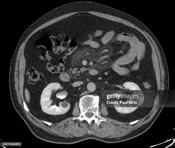
ct findings in sclerosing mesenteritis (panniculitis), coronal view - submucosa stock pictures, royalty-free photos & imagesCT Findings in Sclerosing Mesenteritis (Panniculitis), coronal view
ct findings in sclerosing mesenteritis (panniculitis), axial view - submucosa stock pictures, royalty-free photos & imagesCT Findings in Sclerosing Mesenteritis (Panniculitis), axial viewMedical X-ray, Cut Out, Colon
colorectal cancer (crc), bowel cancer, colon cancer, or rectal cancer seen on sagital t2 mri image - submucosa stock pictures, royalty-free photos & imagesColorectal cancer (CRC), bowel cancer, colon cancer, or rectal cancer seen on sagital T2 MRI imageColorectal Cancer, Cancer Cell, Cancer - Illness
large intestine (colon), layers (or tunics), 10x. shows: mucosa, submucosa, muscularis (smooth muscle - longitudinal and circular layers), serosa, goblet cells (red). - submucosa stock pictures, royalty-free photos & imagesLARGE INTESTINE (COLON), LAYERS (OR TUNICS), 10X. Shows: mucosa, submucosa, muscularis (smooth muscle - longitudinal and circular layers), serosa, goblet cells (red).
esophagus & part of the trachea, cross section, 5x. shows: lumen, mucosa, submucosa, muscularis (smooth muscle), adventitia. trachea: hyaline cartilage, mucous membrane. - submucosa stock pictures, royalty-free photos & imagesESOPHAGUS & PART OF THE TRACHEA, CROSS SECTION, 5X. Shows: lumen, mucosa, submucosa, muscularis (smooth muscle), adventitia. Trachea: hyaline cartilage, mucous membrane.
trachea (windpipe) & esophagus, 2.5x at 35mm. respiratory & digestive systems. trachea shows: lumen, mucosa, cartilage (support), adventia esophagus shows: lumen, mucosa, submucosa, muscularis, adventitia. cross-section. - submucosa stock pictures, royalty-free photos & imagesTrachea (windpipe) & esophagus, 2.5x at 35mm. Respiratory & Digestive Systems. Trachea shows: lumen, mucosa, cartilage (support), adventia Esophagus shows: lumen, mucosa, submucosa, muscularis, adventitia. Cross-section.
small intestine (ileum) with villi and layers (or tunics), 25x shows: mucosa, submucosa, muscularis (circular and longitudinal layers), serosa, intestinal glands, and goblet cells. cross section. - submucosa stock pictures, royalty-free photos & imagesSMALL INTESTINE (ILEUM) WITH VILLI AND LAYERS (OR TUNICS), 25X Shows: mucosa, submucosa, muscularis (circular and longitudinal layers), serosa, intestinal glands, and goblet cells. Cross section.
small intestine with villi and layers (or tunics) 25x. shows: villi, simple columnar epithelium, goblet cells with mucous, submucosa, muscularis (smooth muscle--circular and longitudinal layers) serosa. - submucosa stock pictures, royalty-free photos & imagesSMALL INTESTINE WITH VILLI AND LAYERS (OR TUNICS) 25X. Shows: villi, simple columnar epithelium, goblet cells with mucous, submucosa, muscularis (smooth muscle--circular and longitudinal layers) serosa.
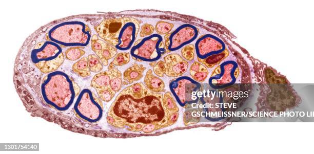
peripheral nerve, tem - submucosa stock pictures, royalty-free photos & imagesPeripheral nerve, TEMFiber
villi.ileum.shows simple columnar epithelium,goblet cells, striated border,submucosa,serosa,25x muscularis - submucosa stock pictures, royalty-free photos & imagesVILLI.ILEUM.SHOWS SIMPLE COLUMNAR EPITHELIUM,GOBLET CELLS, STRIATED BORDER,SUBMUCOSA,SEROSA,25X MUSCULARIS
villi,small intestine.c.s.5x villi,mucosa,submucosa,musculari s, smooth muscle,serosa v - submucosa stock pictures, royalty-free photos & imagesVILLI,SMALL INTESTINE.C.S.5X VILLI,MUCOSA,SUBMUCOSA,MUSCULARI S, SMOOTH MUSCLE,SEROSA V
small intestine human.fetal,cross section.villi, mucosa, submucosa 2.5x circular/longt. muscle layers,serosa long-longitudinal - submucosa stock pictures, royalty-free photos & imagesSMALL INTESTINE HUMAN.FETAL,CROSS SECTION.VILLI, MUCOSA, SUBMUCOSA 2.5X CIRCULAR/LONGT. MUSCLE LAYERS,SEROSA LONG-LONGITUDINALPeople, Cross Section
large intestine - mucosa, submucosa, muscularis, serosa. h - submucosa stock pictures, royalty-free photos & imagesLARGE INTESTINE - MUCOSA, SUBMUCOSA, MUSCULARIS, SEROSA. H
small intestine showing layers---mucosa, submucosa, muscular layer, serosa and simple columnar epithelium with goblet cells, 25x - submucosa stock pictures, royalty-free photos & imagesSMALL INTESTINE showing LAYERS---MUCOSA, SUBMUCOSA, MUSCULAR LAYER, SEROSA and SIMPLE COLUMNAR EPITHELIUM with GOBLET CELLS, 25X
microscopic image of stomach wall cross section - submucosa stock pictures, royalty-free photos & imagesMicroscopic image of stomach wall cross sectionMicroscope
microscopic image of stomach wall cross section - submucosa stock pictures, royalty-free photos & imagesMicroscopic image of stomach wall cross section
microscopic image of stomach wall cross section - submucosa stock pictures, royalty-free photos & imagesMicroscopic image of stomach wall cross section
microscopic image of stomach wall cross section - submucosa stock pictures, royalty-free photos & imagesMicroscopic image of stomach wall cross sectionMicroscope
microscopic image of stomach wall cross section - submucosa stock pictures, royalty-free photos & imagesMicroscopic image of stomach wall cross sectionStomach, Microscope, Cancer - Illness
Site of action of drugs in the treatment of cystic fibrosis. Bronchial wall seen in section with the bronchial mucosa consisting of hair cells, with...Cystic fibrosis