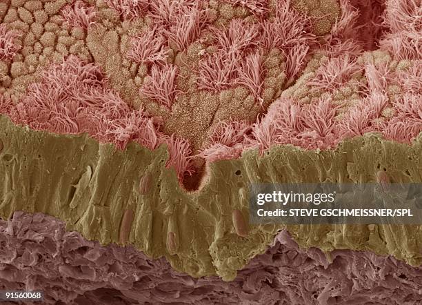
trachea mucous membrane, sem - mucus membrane stock pictures, royalty-free photos & imagesTrachea mucous membrane, SEMMucosal Membrane, SEM
trachea mucous membrane - mucus membrane stock illustrationsTrachea mucous membraneSEM, Mucosal Membrane
microscopic view of bacteria on textured membrane in vibrant 3d visualization - mucus membrane stock pictures, royalty-free photos & imagesMicroscopic View of Bacteria on Textured Membrane in Vibrant 3D VisualizationMucosal Membrane, Antibiotic Resistant
trachea lining, colored scanning electron micrograph - mucus membrane stock pictures, royalty-free photos & imagesTrachea lining, colored scanning electron micrographMucosal Membrane, SEM
rhinosporidium seeberi parasite, illustration - mucus membrane stock illustrationsRhinosporidium seeberi parasite, illustrationIllness, Endangered Species, Parasitic
trachea mucous membrane - mucus membrane stock illustrationsTrachea mucous membraneWineglass, Mucus, Human Gland
trachea lining, colored scanning electron micrograph - mucus membrane stock pictures, royalty-free photos & imagesTrachea lining, colored scanning electron micrographMucus, Epithelium, Cilium
stomach lining, illustration - mucus membrane stock illustrationsStomach lining, illustrationPeople, Biology, Intestine
stomach mucosa, light micrograph and illustration - mucus membrane stock illustrationsStomach mucosa, light micrograph and illustrationStomach
stomach mucosa, light micrograph and illustration - mucus membrane stock illustrationsStomach mucosa, light micrograph and illustrationIntestine
snail trail - mucus membrane stock pictures, royalty-free photos & imagesSnail TrailAustralia, Snail, Escargot
snail trail - mucus membrane stock pictures, royalty-free photos & imagesSnail TrailSnail, Slimy, Escargot
snail trail - mucus membrane stock pictures, royalty-free photos & imagesSnail TrailSnail, Escargot, Slimy
Mucous membrane, Damp epithelial tissue lining an open cavity of the body; the mucous membrane plays a role in absorption and secretion .Mucous membrane
Nasal cavity, The walls of the nasal cavities are covered with a mucous membrane that produces nasal mucus. The odorous molecules dissolve in the...Nasal cavity
Section of the gastric mucous membrane, Heavily folded, the gastric mucous membrane covers the internal surface of the stomach. The thick mucus that...Section of the gastric mucous membrane
Nasal cavity, The walls of the nasal cavities are covered with a mucous membrane that produces nasal mucus. The odorous molecules dissolve in the...Nasal cavity
Histopathologic changes in protothecosis of the skin and mucous membrane of the nose, caused by the achlorophyllic algal organism Prototheca...P. Wickerhamii
Illustration of colon cancer. The cancer develops on the colon's mucus membrane. In 70% of cases, the tumor develops in the sigmoid colon....Cancer of the colon, drawingColonoscopy, Colorectal Cancer, Cancer - Illness
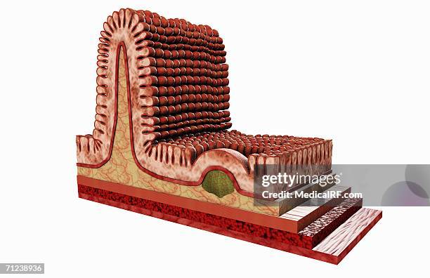
a conceptual visualization of the villi of the small intestine. - mucus membrane stock illustrationsA conceptual visualization of the villi of the small intestine.
illustration showing a conceptual visualization of the villi of the small intestine. - mucus membrane stock illustrationsIllustration showing a conceptual visualization of the villi of the small intestine.
this image depicts a conceptual visualization of the villi of the small intestine. - mucus membrane stock illustrationsThis image depicts a conceptual visualization of the villi of the small intestine.
Illustration of the ciliated cells of the sinus. These cells drain mucus that traps external particles. In the case of chronic sinusitis, the...Sinusitis, Drawing
Illustration of the ciliated cells of the sinus. These cells drain mucus that traps external particles. In the case of sinusitis, the ciliated cells...Sinusitis, Drawing
Illustration of the ciliated cells of the sinus. These cells drain mucus that traps external particles.Sinus, Drawing
Cross-section illustration of the left maxillary sinus, showing the mucous membrane and cilia.Sinus, Drawing
Cross-section illustration of the left maxillary sinus, showing the mucous membrane and cilia.Sinus, DrawingMucosa, Anatomy, Nose