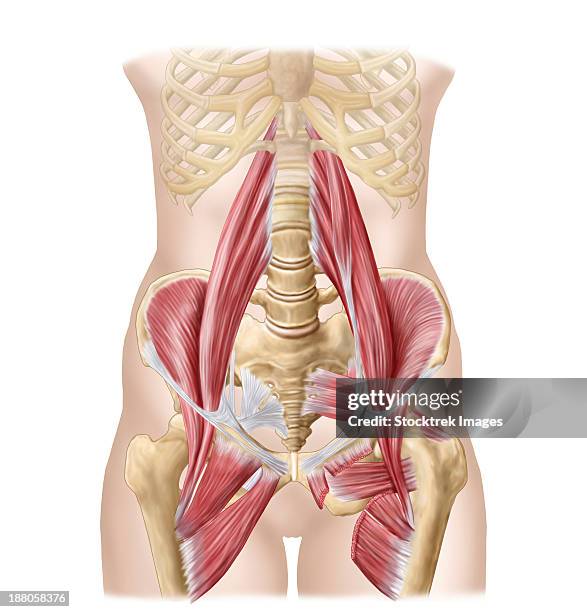
anatomy of iliopsoa, often referred to as the dorsal hip muscles. these muscles are distinct in the abdomen. - iliopsoas stock illustrationsAnatomy of iliopsoa, often referred to as the dorsal hip muscles. These muscles are distinct in the abdomen.Hip - Body Part, Anatomy
anatomy of iliopsoa, often referred to as the dorsal hip muscles. these muscles are distinct in the abdomen. - iliopsoas stock illustrationsAnatomy of iliopsoa, often referred to as the dorsal hip muscles. These muscles are distinct in the abdomen.
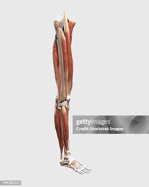
medical illustration of human leg muscles, bones and joints. - iliopsoas stock illustrationsMedical illustration of human leg muscles, bones and joints.
The human body with superimposed colored plates, by Julien Bougle, circa 1899.The human body with superimposed colored plates, by Julien Bougle, circa 1899.
woman doctor adjusting patients knee - iliopsoas stock pictures, royalty-free photos & imagesWoman doctor adjusting patients knee
doctor doing a knee to chest assessment - iliopsoas stock pictures, royalty-free photos & imagesDoctor doing a knee to chest assessment
woman doctor evaluating patients hip - iliopsoas stock pictures, royalty-free photos & imagesWoman doctor evaluating patients hip
woman doctor evaluating patients knee - iliopsoas stock pictures, royalty-free photos & imagesWoman doctor evaluating patients knee
male muscle anatomy of the human legs, anterior view. - iliopsoas stock illustrationsMale muscle anatomy of the human legs, anterior view.
male muscle anatomy of the human legs, anterior view. - iliopsoas stock illustrationsMale muscle anatomy of the human legs, anterior view.
medical illustration of human leg muscles, four side views. - iliopsoas stock illustrationsMedical illustration of human leg muscles, four side views.Anatomy, Low Section, Digitally Generated Image
diagram illustrating muscle groups on front of human legs. - iliopsoas stock illustrationsDiagram illustrating muscle groups on front of human legs.Low Section
Illustration from 'Atlas and Epitome of Traumatic Fractures and Dislocations' , 1902. Fig 1 Muscle preparation to explain the flexion of the upper...Typical Deformity In Fracture Of The Middle Of The Femur
Axial Section. Cf. Image 0212906 For The Numbers. 1. Rectus Abdominis Muscle. 2. Inguinal Canal And Spermatic Cord. 3. Femoral Artery. 4. Femoral...Pelvis, Scan
Axial Section. Cf. Image 0212906 For The Numbers. 1. Rectus Abdominis Muscle. 2. Inguinal Canal And Spermatic Cord. 3. Femoral Artery. 4. Femoral...Pelvis, Scan
Axial Section. 1. Rectus Abdominis Muscle. 2. Inguinal Canal And Spermatic Cord. 3. Femoral Artery. 4. Femoral Vein. 5. Iliopsoas Muscle. 6. Gluteus...Pelvis, Scan
The Anterior View Of The Muscles Of The Human Right Leg.The Anterior View Of The Muscles Of The Human Right Leg.Anatomy, Leg, Front View
low section front view of human limbs, hip and muscular system with arteries and veins. - iliopsoas stock illustrationsLow section front view of human limbs, hip and muscular system with arteries and veins.
low section front view of human limbs, hip and muscular system with arteries and veins. - iliopsoas stock illustrationsLow section front view of human limbs, hip and muscular system with arteries and veins.
low section front view of human limbs, hip and muscular system, on white background. - iliopsoas stock illustrationsLow section front view of human limbs, hip and muscular system, on white background.
digital illustration of the anterior muscles of the leg (no labels). - iliopsoas stock illustrationsDigital illustration of the anterior muscles of the leg (no labels).
digital illustration of the posterior muscles of the leg. - iliopsoas stock illustrationsDigital illustration of the posterior muscles of the leg.Gastrocnemius, Anatomy
digital illustration of the posterior muscles of the leg (no labels). - iliopsoas stock illustrationsDigital illustration of the posterior muscles of the leg (no labels).
a diagram depicting the muscles in the upper body, from the backside. - iliopsoas stock illustrationsA diagram depicting the muscles in the upper body, from the backside.
lateral and posterior view of female hip and leg muscles. - iliopsoas stock illustrationsLateral and posterior view of female hip and leg muscles.
lateral and posterior view of female hip and leg muscles. - iliopsoas stock illustrationsLateral and posterior view of female hip and leg muscles.
rear view of female hip and leg muscles on white background. - iliopsoas stock illustrationsRear view of female hip and leg muscles on white background.
rear view of female hip and leg muscles, with labels. - iliopsoas stock illustrationsRear view of female hip and leg muscles, with labels.
female chest and abdomen muslces, pink x-ray view. - iliopsoas stock illustrationsFemale chest and abdomen muslces, pink x-ray view.