h&e stain, light microscopy, malignant melanoma of skin - h e stain stock pictures, royalty-free photos & imagesH&E stain, light microscopy, malignant melanoma of skinMelanoma, Microscope, Skin Cancer
h&e stain, light microscopy, multiple myeloma - h e stain stock pictures, royalty-free photos & imagesH&E stain, light microscopy, multiple myelomaMultiple Myeloma, Human Cell, Healthcare And Medicine
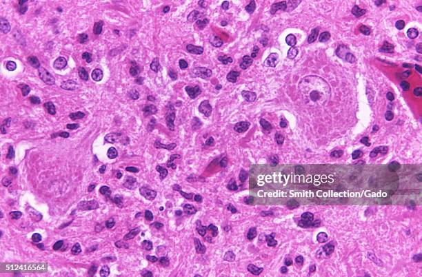
Cellular changes associated with rabies encephalitis, revealed in the photomicrograph using H and E staining, 1971. Image courtesy Centers for...Rabies Encephalitis
Samples of lung tissue from D M, case number 81 in Elmes' work with McCaughey and Wade to produce the first detailed UK report of the relationship...Slides of lung tissue from Dr P C Elmes slide cabinet, c 1970.
Samples of lung tissue from D M, case number 81 in Elmes' work with McCaughey and Wade to produce the first detailed UK report of the relationship...Slides of lung tissue from Dr P C Elmes slide cabinet, c 1970.
Samples of lung tissue from G C, case number 48 in Elmes' work with McCaughey and Wade to produce the first detailed UK report of the relationship...Slides of lung tissue from Dr P C Elmes slide cabinet, c 1965.
Samples of lung tissue from R A, case number 86 in Elmes' work with McCaughey and Wade to produce the first detailed UK report of the relationship...Slides of lung tissue from Dr P C Elmes slide cabinet, c 1973.
Samples of lung tissue from J L S, case number 84 in Elmes' work with McCaughey and Wade to produce the first detailed UK report of the relationship...Slides of lung tissue from Dr P C Elmes slide cabinet, c 1969.
Samples of lung tissue from G C, case number 48 in Elmes' work with McCaughey and Wade to produce the first detailed UK report of the relationship...Slides of lung tissue from Dr P C Elmes slide cabinet, c 1965.
creutzfeldt-jakob disease brain, 68yo man, following flu-like syndrome. h&e stain/100x - h e stain stock pictures, royalty-free photos & imagescreutzfeldt-jakob disease brain, 68yo man, following flu-like syndrome. h&e stain/100xIllness
siderosis. hematite miners lung with collections of dark dust particles (macules). h&e stain. 26x: - h e stain stock pictures, royalty-free photos & imagessiderosis. hematite miners lung with collections of dark dust particles (macules). h&e stain. 26x:
These encephalitic changes found in a 2 week old chicken brain were due to the fungal organism Ochroconis gallopavum, formerly Dactylaria gallopava,...Ochroconis Gallopavum
Photomicrograph detecting the presence of hemosiderin, stained blue, within diffuse bone marrow, 1972. Hemosiderin, revealed here using PAS stain and...Diffuse Bone Marrow
Photomicrograph detecting the presence of hemosiderin, stained blue, within diffuse bone marrow, 1972. Hemosiderin, revealed here using PAS stain and...Diffuse Bone Marrow
This pinta lesion, stained using the hematoxylin-eosin technique, is caused by the bacterium Treponema carateum, magnification 100X, 1970. This...Treponema Carateum
Photomicrograph of lung tissue revealing Pneumonia Alba, which is caused by congenital syphilis, 1971. An alveolar duct is seen in the center. The...Pneumonia AlbaTreponema Pallidum
Photomicrograph of a condyloma latum lesion, indicative of secondary syphilis, 1971. Pathognomonic findings seen here include acanthosis, broadening...Condyloma Latum Lesion
Photomicrographic study using H&E stain, of an early pinta, a skin lesion caused by the bacterium Treponema carateum, 1970. Note the contrast between...Treponema Carateum
Photomicrograph of osteochondritis revealing the cytoarchitectural changes due to congenital syphilis, 1971. There is marked irregularity in the...Courtesy Cdc/Susan Lindsley
Photomicrograph revealing cytoarchitectural changes seen in congenital syphilis of the placenta, 1971. The chorionic villi are enlarged, and contain...Connective Tissue Proliferation
Photomicrograph of Escherichia coli, bacteria using Gram-stain technique, 1979. E coli O157:H7 is one of hundreds of strains of the bacterium...Escherichia ColiE. coli
Photomicrograph of hematoxylin-eosin stained brain tissue from a rabies encephalitis patient. Histopathologic brain tissue from a rabies patient...Rabies encephalitis patientRabies
Photomicrograph of H&E stained brain tissue from a rabies encephalitis patient. Histopathologic brain tissue from a rabies patient displaying the...Rabies encephalitis patient
Photomicrograph of the cervical spinal cord affected by polio type-III virus. Histopathologic changes seen in a section of cervical spinal cord...Polio type-III virusPolio, USA
Necrosis of lymph node due to anthrax. 33 year old white male who died of inhalation anthrax, photomicrograph demonstrating necrotic lymph node found...Necrosis Of Lymph Node Due to AnthraxLymphatic System
h&e stain, light microscopy, paget disease of bone - h e stain stock pictures, royalty-free photos & imagesH&E stain, light microscopy, Paget disease of bone
h&e stain, light microscopy, abundant collagen in a gardner fibroma - h e stain stock pictures, royalty-free photos & imagesH&E stain, light microscopy, abundant collagen in a Gardner fibromaCollagen
h&e stain, light microscopy, healing fracture callus with exuberant cartilage and new bone formation - h e stain stock pictures, royalty-free photos & imagesH&E stain, light microscopy, healing fracture callus with exuberant cartilage and new bone formation
h&e stain, light microscopy, rheumatoid arthritis of joint showing pannus - h e stain stock pictures, royalty-free photos & imagesH&E stain, light microscopy, rheumatoid arthritis of joint showing pannusRheumatoid Arthritis
h&e stain, light microscopy, normal hair follicle - h e stain stock pictures, royalty-free photos & imagesH&E stain, light microscopy, normal hair follicle
h&e stain, light microscopy, rheumatoid arthritis of joint showing inflammation and hyperplasia - h e stain stock pictures, royalty-free photos & imagesH&E stain, light microscopy, rheumatoid arthritis of joint showing inflammation and hyperplasiaRheumatoid Arthritis, Hyperplasia, Magnification
h&e stain, light microscopy, normal skin of scalp - h e stain stock pictures, royalty-free photos & imagesH&E stain, light microscopy, normal skin of scalpMicroscope, Human Cell, Human Scalp
h&e stain, light microscopy, osteoporosis, bone - h e stain stock pictures, royalty-free photos & imagesH&E stain, light microscopy, osteoporosis, bone
histology micrograph of renal failure,showing interstitial nephritis in tissue of kidney with h&e stain - h e stain stock pictures, royalty-free photos & imagesHistology micrograph of renal failure,showing interstitial nephritis in tissue of kidney with H&E stain
histology micrograph of renal failure,showing interstitial nephritis in tissue of kidney with h&e stain - h e stain stock pictures, royalty-free photos & imagesHistology micrograph of renal failure,showing interstitial nephritis in tissue of kidney with H&E stain
histology micrograph of renal failure,showing interstitial nephritis in tissue of kidney with h&e stain - h e stain stock pictures, royalty-free photos & imagesHistology micrograph of renal failure,showing interstitial nephritis in tissue of kidney with H&E stain
histology micrograph of renal failure,showing interstitial nephritis in tissue of kidney with h&e stain - h e stain stock pictures, royalty-free photos & imagesHistology micrograph of renal failure,showing interstitial nephritis in tissue of kidney with H&E stainInflammation, Tissue - Anatomy, Healthcare And Medicine
histology micrograph of renal failure,showing interstitial nephritis in tissue of kidney with h&e stain - h e stain stock pictures, royalty-free photos & imagesHistology micrograph of renal failure,showing interstitial nephritis in tissue of kidney with H&E stain
Photomicrograph of a tissue specimen from an actinomycotic mycetoma, stained with hematoxylin and eosin , at 900X magnification, 1965....Nocardia Madurae Tissue Photomicrograph
Photomicrograph of a hematoxylin and eosin stained tissue section from an actinomycotic mycetoma, showing histopathologic changes due to Actinomadura...Actinomadura Pelletieri Histopathology
caucasian female professor lectures on h&e stain analysis - h e stain stock pictures, royalty-free photos & imagesCaucasian Female Professor Lectures on H&E Stain AnalysisPresentation - Speech, Research, Professor
At the church is a stained glass portrait of Rachel Bassette Noel <cq>. It is second from right. A memorial service celebrating and honoring the life...(HR) ABOVE: At the church is a stained glass portrait of Rachel Bassette Noel <cq>. It is second from right. A memorial service celebrating and honoring the life of Rachel Bassette Noel <cq> was held at the Shorter Community A.M.E Church today March 2, 20
slide staining set for histopathology (h&e stain) - h e stain stock pictures, royalty-free photos & imagesslide staining set for histopathology (H&E stain)
Influenza Virus Colorized Tem, Negative Stained. Surface Glycoproteins Are Clearly Visible. Influenzavirus > Orthomyxoviridae > Rna Virus > Virus....Influenza Virus, TemVirus, Flu Virus, Microscope
sebaceous glands and hair follicle of human scalp (magnification x25) h & e stain. showing hair follicle, hair, epidermis and dermis and arrector pili muscle - h e stain stock pictures, royalty-free photos & imagesSebaceous Glands and Hair Follicle of Human Scalp (Magnification x25) H & E stain. Showing hair follicle, hair, epidermis and dermis and arrector pili muscleHair Follicle
cardiac muscle with intercalated discs, human, (magnification x250). showing intercalated discs, striations, nucle and syncytium (branching, interconnected network of cells). involuntary muscle. h & e stain. - h e stain stock pictures, royalty-free photos & imagesCardiac Muscle with Intercalated Discs, Human, (Magnification x250). Showing intercalated discs, striations, nucle and syncytium (branching, interconnected network of cells). Involuntary muscle. H & E stain.Microscope, Tissue - Anatomy, Human Tissue
capillary with red blood cells in rouleaux (magnification x250) h & e stain. red blood cells (erythrocytes) can form stacks called rouleaux which can form and dissociate repeatedly, facilitating movement through the small capillaries. also shows capillary - h e stain stock pictures, royalty-free photos & imagesCapillary with Red Blood Cells in Rouleaux (Magnification x250) H & E stain. Red Blood Cells (Erythrocytes) can form stacks called rouleaux which can form and dissociate repeatedly, facilitating movement through the small capillaries. Also shows capillaryCapillary - Body Part, Blood Vessel, Connection
hair papilla, bulb and follicle of human scalp (magnification x50) h & e stain: the base of the hair follicle is where growth takes place. the papilla shows a capillary with red blood cells. - h e stain stock pictures, royalty-free photos & imagesHair Papilla, Bulb and Follicle of Human Scalp (Magnification x50) H & E stain: The base of the hair follicle is where growth takes place. The papilla shows a capillary with red blood cells.Hair Follicle
normal artery in cross section (magnification x100) h & e stain. three distinct layers or tunics: tunica interna (intima), tunica media, tunica externa (adventitia). also shows endothelium and lumen with red and white blood cells. - h e stain stock pictures, royalty-free photos & imagesNormal Artery in Cross Section (Magnification x100) H & E stain. Three distinct layers or tunics: tunica interna (intima), tunica media, tunica externa (adventitia). Also shows endothelium and lumen with red and white blood cells.Thyroid Gland, Tissue - Anatomy, Photography
sebaceous glands and hair follicle of human scalp (magnification x25) h & e stain. sebaceous glands, hair follicle and a hair in the follicle. - h e stain stock pictures, royalty-free photos & imagesSebaceous Glands and Hair Follicle of Human Scalp (Magnification x25) H & E stain. Sebaceous glands, hair follicle and a hair in the follicle.Magnification, Hair Follicle
sebaceous glands and hair follicle of human scalp (magnification x25) h & e stain. sebaceous glands (clear cells that contain oil), hair follicle, hair, epidermis and dermis. - h e stain stock pictures, royalty-free photos & imagesSebaceous Glands and Hair Follicle of Human Scalp (Magnification x25) H & E stain. Sebaceous glands (clear cells that contain oil), hair follicle, hair, epidermis and dermis.
smooth muscle of human bladder (magnification x 100) h & e stain. nonstriated muscle showing nuclei, cytoplasm and tapered structure of cells. - h e stain stock pictures, royalty-free photos & imagesSmooth Muscle of Human Bladder (Magnification x 100) H & E stain. Nonstriated muscle showing nuclei, cytoplasm and tapered structure of cells.
smooth muscle of human bladder (magnification x 250) h & e stain. nonstriated muscle showing nuclei, cytoplasm and tapered structure of cells. - h e stain stock pictures, royalty-free photos & imagesSmooth Muscle of Human Bladder (Magnification x 250) H & E stain. Nonstriated muscle showing nuclei, cytoplasm and tapered structure of cells.
stratified squamous epithelium of human vagina (magnification x 50) h & e stain. non-keratinized. cells become very flat near surface. - h e stain stock pictures, royalty-free photos & imagesStratified Squamous Epithelium of Human Vagina (Magnification x 50) H & E stain. Non-keratinized. Cells become very flat near surface.Tissue - Anatomy
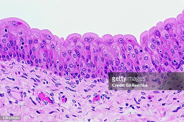
transitional epithelium of human bladder (magnification x100). h & e stain. cells near the surface are the pear-shaped basement membrane, with supporting connective tissue below - h e stain stock pictures, royalty-free photos & imagesTransitional Epithelium of Human Bladder (Magnification x100). H & E stain. Cells near the surface are the pear-shaped basement membrane, with supporting connective tissue belowEpithelium, Tissue - Anatomy, Bladder
transitional epithelium of human bladder (magnification x100). h & e stain. cells near the surface are the pear-shaped basement membrane, with supporting connective tissue below - h e stain stock pictures, royalty-free photos & imagesTransitional Epithelium of Human Bladder (Magnification x100). H & E stain. Cells near the surface are the pear-shaped basement membrane, with supporting connective tissue below
Case with Design of Poppies, Edo period , Japan, Case: powdered gold and gold and colored lacquer on black lacquer with pewter and ceramic inlays;...Case (Inr_) with Design of Poppies
Case with Design of Grasshopper on Stalk of Flowering Lily, Edo period , 18th-19th century, Japan, Case: Powdered gold on black lacquer with...Case (Inr_) with Design of Grasshopper on Stalk of Flowering Lily